42 areolar tissue diagram
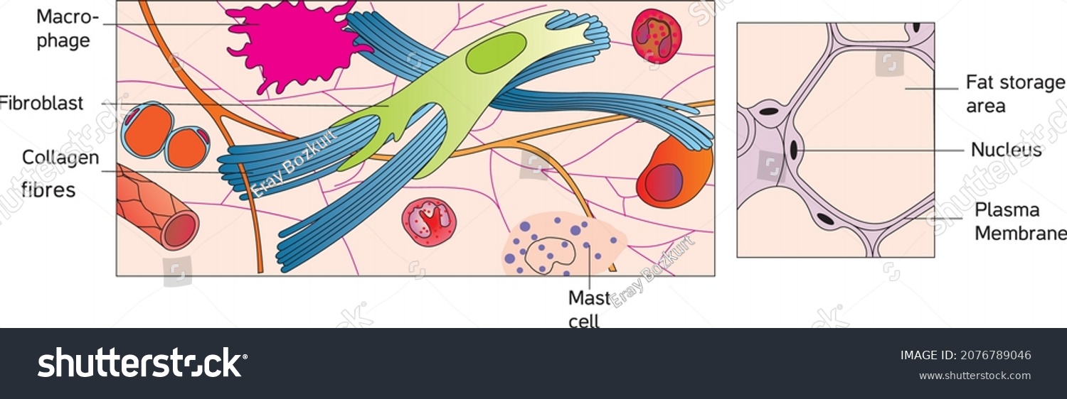
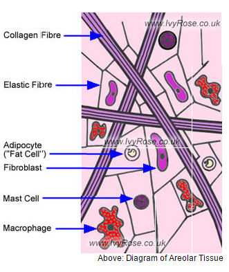
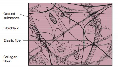
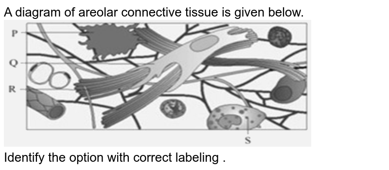
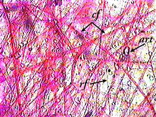
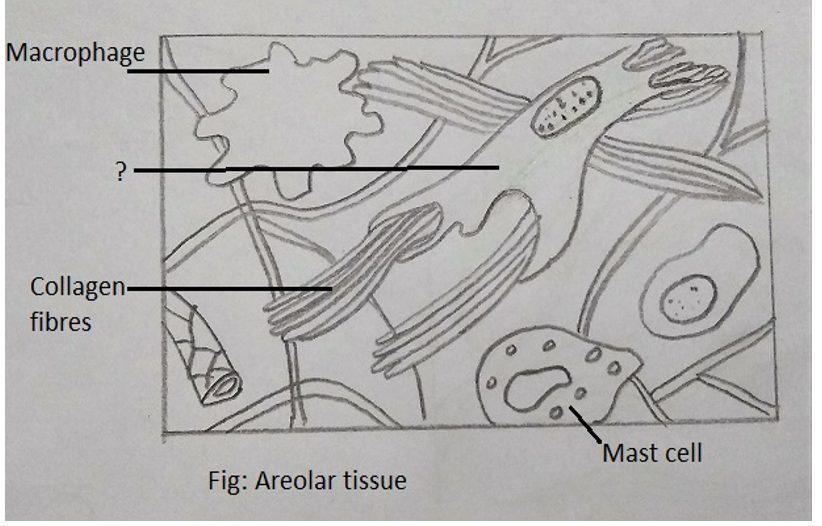
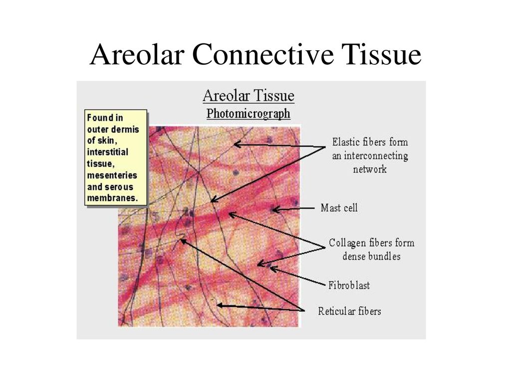
Areolar tissues are widely distributed in the body and primarily function as a packing material between other tissues. Functions of the Areolar Connective Tissue. The areolar connective tissue is a type of connective tissue that is present throughout the human body. It provides support and helps to protect organs, muscles, and many other tissues. Areolar connective tissue is a loosely arranged connective tissue that is widely distributed in the Body and contains collagen fibres, reticular fibres and a few elastic fibres embedded in a thin, almost fluid-like ground substance.
Loose Connective Tissue | histologyolm.stevegallik.org Loose connective tissue - Wikipedia Classification of Tissues - Cardiovascular Technician 1 ... Loose Connective Tissue Stock Photo - Download Image Now ... Connective Tissue Types and Examples Connective Tissue - WhatMaster Loose Connective Tissue: Function, Structure, Types and ... Loose Connective Tissue - Assignment Point Histology - Medicine Medi7111 with Dr. Tammy Snith at ... Magnified Image Of Loose Connective Tissue Stock Photo ...

Areolar tissue diagram
As stated earlier, the areolar tissue is the most widely distributed connective tissue in the body. It is present under the skin as subcutaneous tissue in between and around muscles, nerves and blood vessels in sub-mucosa of gastrointestinal tract and respiratory tract, in the bone marrow, between the lobes and lobules of compound glands and in ... Sep 18, 2021 · Labelled Diagram Of Areolar Connective Tissue. angelo on September 18, 2021. Pin On Anatomy Lab. Integumentary System Diagram Google Search Skin Anatomy Integumentary System Human Integumentary System. Pin On Career. Areolar Connective Tissue Diagram. This diagram shows areolar connective tissue serving as a medium between epithelial tissue and blood vessels. Cellular components (such as fibroblasts) and the ...
Areolar tissue diagram. The human areola (areola mammae, / ə ˈ r iː ə l ə / or / ˌ ær i ˈ oʊ l ə /) is the pigmented area on the breast around the nipple.Areola, more generally, is a small circular area on the body with a different histology from the surrounding tissue, or other small circular areas such as an inflamed region of skin.. The mature human female nipple has several small openings arranged ... Name the tissue present in the diagram. A) Bone B) Areolar Tissue C) Adipose Tissue D) Cartilage Tissue Connective Tissue Types. Loose connective tissue types. Areolar tissue. Most widely distributed connective tissue. Soft, pliable tissue like "cobwebs" Functions as a universal packing tissue and "glue" to hold organs in place. Layer of areolar tissue called . lamina . propria. underlies all membranes. All fiber types form a loose network Also includes what appears to be a draft of On Some Mechanical Functions of Areolar Tissues, which appeared in Amer. J. of the Medical Sciences, vol. XXII (Phila., Carey, Lea & Blanchard, 1838). Persons mentioned in the journal text include William H. Goode, Dr. Dalton (John Dalton?), Dr. Ritchie (William Ritchie?). 리뷰 사용자-기여... Cookie Notice OCLC 웹사이트는 사용자의 경험을 개선하기 위해 사용자의 장치에 쿠키를 저장합니다. 자세한...
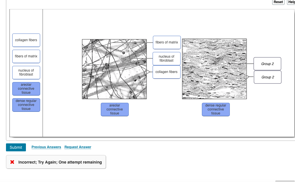
The lung root is formed by: the bronchus, the pulmonary artery and veins, the bronchial arteries and veins, the pulmonary plexuses of nerves, lymphatic vessels, bronchial lymph glands, and areolar tissue, all of which are enclosed by a layer of the pleura, which is a thin smooth layer of protective tissue. More specifically, a... ADVERTISEMENTS: The following points highlight the ten main varieties of connective tissues of human body. They are: 1. Areolar Tissue 2. Adipose Tissue 3. White Fibrous Tissue 4. Yellow Elastic Tissue 5. Reticular Tissue 6. Blood and Haemopoietic Tissue 7. Cartilage 8. Jelly-Like Connective Tissue 9. Osseous Tissue or Bone 10. Reticulo-Endothelial Tissue. 1. Areolar […] Areolar Tissue Diagram. Areolar Connective Tissue Definition Areolar connective tissue is a loosely arranged. To bind parts together is the primary function of areolar tissue. Other functions are to provide strength, elasticity, support to the parts where this tissue is present. Figure Diagram of areolar tissue as it appears in microscopic section. 이 세트의 단어 (11) Compact bone High strength, low flexibility tissue in the cortex of bones Areolar tissue Stretchy, cobweb-like tissue that underlies most epithelia and wraps organs, blood vessels, and nerves Dense regular fibrous connective tissue The structural tissue of tendons and ligaments; transmits force in one...
Draw a labelled diagram of areolar connective tissue. structural organisation in animals; class-11; Share It On Facebook Twitter Email. 1 Answer +1 vote . answered Feb 5, 2020 by KumariJuly (53.6k points) selected Feb 5, 2020 by Ritik01 . Best answer. Diagram of areolar connective tissue ... areolar tissue diagram - Google Search. Denver Jaque. Debut Invitation. Invitation Cards. Tissue Biology. Human Tissue. Tissue Types. Drawing Sketches. Drawings. Body Tissues ... cell and tissue studies, and an overview of all the body systems. Intended as a survey course for certain allied health and social service programs, and as a general ... cord-like tissue connecting two larger parts of an anatomical structure - the tissue in the bodies of animals - a soft thin (usually translucent) paper , - a strong tissue paper that burns evenly and is sufficiently porous to control the burning of the tobacco in a cigarette - tissue paper suitable for use on the face - a piece... Dec 13, 2018 · Areolar Connective Tissue Diagram. Description of matrix, fibres and cells has already been given in the general structure of connective tissue. Location: As stated earlier, the areolar tissue is the . Areolar connective tissue has no obvious structure, like layers or rows of cells. You might think that this would make it harder to identify.
In the given diagram of areolar tissue, identify a phagocytic cell : 1. A 2. B 3. C 4. D Structural Organisation in Animals Zoology - Mini Question Bank Practice questions, MCQs, Past Year Questions (PYQs), NCERT Questions, Question Bank, Class 11 and Class 12 Questions, NCERT Exemplar Questions and PDF Questions with answers, solutions, explanations, NCERT reference and difficulty level
Here are a number of highest rated Computer Function Diagram pictures on internet. We identified it from trustworthy source. Its submitted by management in the best field. We believe this nice of Computer Function Diagram graphic could possibly be the most trending subject later than we allowance it in google plus or facebook.
Learn areolar tissue with free interactive flashcards. Choose from 88 different sets of areolar tissue flashcards on Quizlet.
Aug 23, 2018 · Areolar connective tissues hold organs in place and attaches epithelial tissue to other underlying tissues. It also serves as a reservoir of. In this lesson, you'll explore what areolar connective tissue is, what elements Simple Cuboidal Epithelium: Location, Structure & Function.Dec 11, · Diagram Of Areolar Tissue.
The skin lies on the subcutaneous tissue ("superficial fascia"), a layer of fatty areolar tissue that overlies the more densely fibrous fascia. It should be remembered that fat is liquid, or nearly so, at body temperature. The subcutaneous tissue serves as a depot for fat storage and aids in preventing loss of heat. When a... Basic Human Anatomy - O'Rahilly, Müller, Carpenter & Swenson Etymology Glossary Table of Contents Copyright...
Areolar tissue (in skin) ... Dense irregular fibrous connective tissue (in skin) The structural tissue of skin; can be tanned to produce leather. Cilia Motorized "hairs" used to push fluids such as mucus Goblet cell Cells that produce mucus Capillaries and lacteals Vessels that receive the nutrients absorbed by the intestinal...
Connective tissue is composed primarily of two elements:and a . The types of cells found in connective tissue vary depending on the type of tissue they support. For example, red and white blood cells are found in blood, which is a fluid connective tissue. Adipocytes are fat cells found in adipose tissue, or fat. And fibroblasts...
Complete the diagram: Cell -> ____ -> Organs -> ____ Answer: tissue, system 4. Connetive tissue is classifed as loose or ___ ; muscle tissue is classifed as smotth... secretion (glands) Connective loose or dense special connective tissues include cartilage, bone, blood, and lymph protection (areolar); storage(adipose)...
Areolar Tissue. Areolar tissue is the most widely distributed solid connective tissue in the body. Which makes sense because areolar tissue is present under the entire skin. And areolar tissue also supports the epithelium. Areolar tissue contains randomly spread out fibers, fibroblasts, mast cells, and macrophages.
Mucosa = stratified squamous epithelium on the surface with a paler connective tissue lamina propria underneath it and a darker, denser muscularis mucosae below the connective tissue. Submucosa = lighter connective tissue layer. Notice the submucosal gland with its duct extending up to the surface. Muscularis externa = darker...
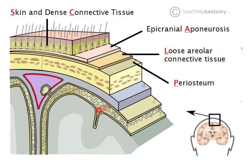
Mar 04, 2019 · Areolar tissue is found in many locations around the body. One important area is the skin (areolar tissue is found in both the dermis and sub-cutaneous layers of the skin - see diagram).The areolar tissue located in the skin binds the outer layers of the skin to the muscles beneath. Areolar connective tissue. wraps: blood vessels nerves glands ...
The Structure Of Areolar Tissue. Areolar Tissue is the areolar connective tissue that consists of a meshwork of collagen, elastic tissue, and reticular fibers - with many connective tissue cells in between the meshwork of fibers. Areolar Connective Tissue Labelled Diagram. The fibers that construct the mesh structure of areolar tissue include ...
Start studying Areolar Tissue. Learn vocabulary, terms, and more with flashcards, games, and other study tools.
Areolar connective tissue is the most abundant of the connective tissues in the body. It is sturdier than adipose tissue but still flexible. Areolar connective tissue is predominantly found in the ...
The effects of progressive formaldehyde fixation on the preservation of tissue antigens. . Bancroft JD and Gamble M. Theory and practice of histological techniques . Churchill Livingstone Elsevier, Philadelphia, PA, 2008. Lester SC, Bose S, Chen YY, Connolly JL, de Baca ME, Fitzgibbons PL, et al. Protocol for the examination of...
How to draw a Diagram of Areolar Tissue in exam is the topic. This is the well labelled diagram of structure of Areolar Tissue. This is a well labelled diagr...
Breast anatomy is complex and intricate, involving various types of tissue, including milk ducts, lymph vessels, the nipple and areola, and other structures. Here's what to know about the many internal and external parts of the breast, their purpose, and the medical conditions that can affect them.
Please Give A Detailed Diagram Of Areolar And Adipose Connective Tissue With Proper Explanation Of Each Component Of It Biology Topperlearning Com Zh17wp88. With Help Of Neat Labelled Diagram Describe The Structure Of Areolar Connective Tissue Brainly In. 33 2b Connective Tissues Loose Fibrous And Cartilage Biology Libretexts.
Click here 👆 to get an answer to your question ️ draw a labeled diagram of areolar tissue suyash97 suyash97 30.04.2019 Biology Secondary School answered Draw a labeled diagram of areolar tissue 2 See answers Advertisement Advertisement SHRUTI2702 SHRUTI2702
Drugs & Diseases > Clinical Procedures Mandibular Plating Updated: Oct 29, 2018 Author: Jesse E Smith, MD, FACS; Chief Editor: Arlen D Meyers, MD, MBA more... 4 Overview The reconstruction of oromandibular defects (mandibular reconstruction) following surgical extirpation of oral cavity carcinoma presents a significant surgical... This site is intended for healthcare professionals English Edition Register Log In News & Perspective Drugs...
Areolar tissue is a loose connective tissue found under the skin, between muscles, bones, around organs, blood vessels, and peritoneum. It is composed of fibers and cells. The matrix of areolar tissues contains two types of fibres i.e. white fibres and yellow fibres. White fibres: They are made up of collagen and give tensile strength to the ...
The sclerotomes are destined to give rise to the vertebrae and other forms of connective tissue in their... connective tissue forming parts of the other organs of the body. Fig. 101. Transverse section of chick embryo... Mesenchymal tissue from somatopleure of a 5 mm human embryo. Mesothelium is shown along lower border of...
A tissue is a group of, in close proximity, organized to perform one or more specific functions. There are four basic tissue types defined by their morphology and function: epithelial tissue, connective tissue, muscle tissue, and nervous tissue.creates protective boundaries and is involved in the diffusion of ions and...
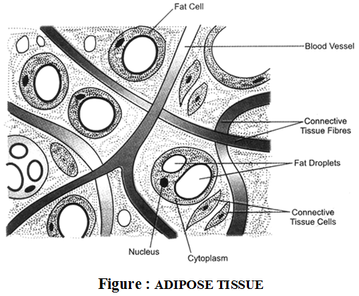
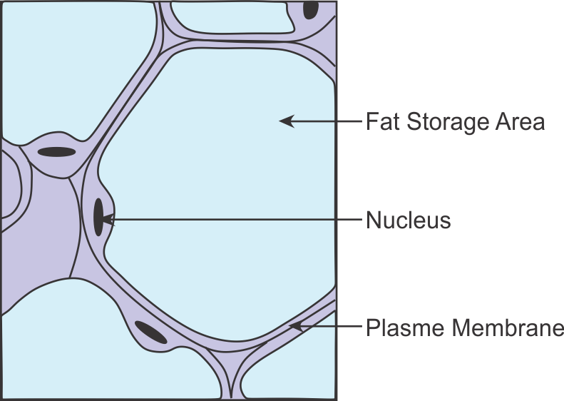
Helps in the repair of tissues. Adipose tissue Found beneath the skin, around the kidneys and other internal organs such as intestines. Cells are filled with fat globules situated in a large central vacuole of a cell, pushing the cytoplasm and nucleus to the periphery. A loose framework of areolar tissue supports these cells.
About Press Copyright Contact us Creators Advertise Developers Terms Privacy Policy & Safety How YouTube works Test new features Press Copyright Contact us Creators ...
Areolar Connective Tissue Diagram. This diagram shows areolar connective tissue serving as a medium between epithelial tissue and blood vessels. Cellular components (such as fibroblasts) and the ...
Sep 18, 2021 · Labelled Diagram Of Areolar Connective Tissue. angelo on September 18, 2021. Pin On Anatomy Lab. Integumentary System Diagram Google Search Skin Anatomy Integumentary System Human Integumentary System. Pin On Career.
As stated earlier, the areolar tissue is the most widely distributed connective tissue in the body. It is present under the skin as subcutaneous tissue in between and around muscles, nerves and blood vessels in sub-mucosa of gastrointestinal tract and respiratory tract, in the bone marrow, between the lobes and lobules of compound glands and in ...














Comments
Post a Comment